


Patients undergoing treatment at the Garmed Clinic undergo a comprehensive laboratory (tests), hardware (ECG, ultrasound, MRI, CT, EEG), and psychodiagnostic (pathopsychological examination, testing) examination. Diagnosis of physical and psychological condition is an important factor in a person’s recovery.
Some examinations can be provided by our clinic’s partners.
Laboratory analyses
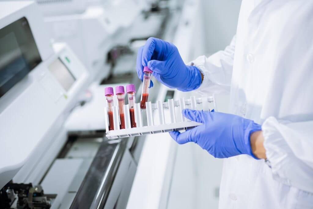
The medical centre “GarMed” conducts a full range of analyses that are necessary for our clients.
Analysis (test) for drugs and alcohol
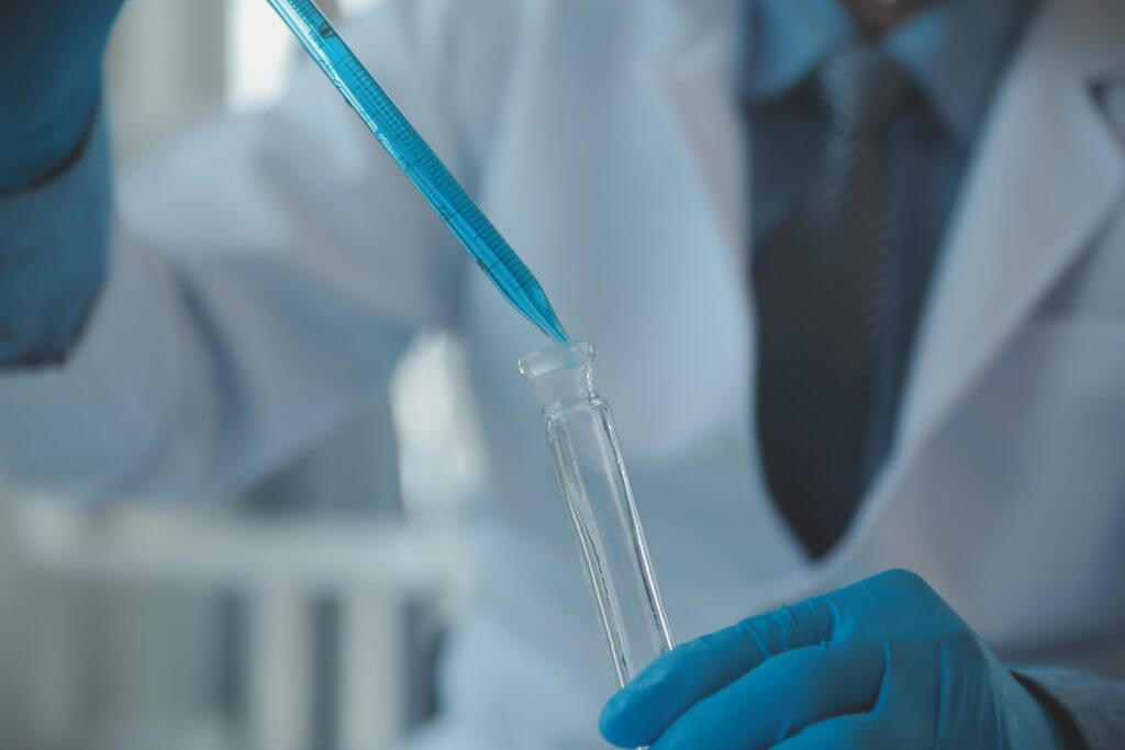
If you need to take a drug and alcohol test or suspect that your loved one is using drugs, you can take a drug and alcohol test and get expert advice at our centre.
Psychodiagnostics

During the course of treatment and rehabilitation, patients undergo diagnostics of their psychological and psycho-emotional state, and with the help of psychological diagnostic methods (pathopsychological examination) and various types of psychological testing, specialists are able to plan and adjust the course of treatment.
Electrocardiogram (ECG)

Electrocardiography (abbreviated as ECG) is a method of graphically recording electrical phenomena that occur in the heart muscle during its activity from the body surface. The curve that reflects the electrical activity of the heart is called an electrocardiogram (ECG). Thus, ECG is a record of potential difference fluctuations that occur in the heart during its excitation.
Electrocardiography is one of the main ways to examine the heart and diagnose diseases of the cardiovascular system. ECG is indispensable in the diagnosis of rhythm and conduction disorders, hypertrophy, and coronary heart disease. This method makes it possible to speak with great accuracy about the localisation of focal myocardial changes, their prevalence, depth and time of occurrence. ECG allows to detect dystrophic and sclerotic processes in the myocardium, electrolyte metabolism disorders arising under the influence of various toxic substances. ECG is widely used for functional examination of the cardiovascular system. The combination of electrocardiographic examination with functional tests helps to detect latent coronary insufficiency, transient rhythm disturbances, and to make a differential diagnosis between functional and organic heart disorders.
In the XIX century, it was discovered through pathology research that the heart has an electrical potential and produces a certain amount of electrical energy during its work. The first ECGs were recorded in 1888 by Gabriel Lippmann, a French physicist who won the 1908 Nobel Prize in Physics, using a mercury capillary electrometer. But this technique did not find practical use. In 1903, the Dutch physiologist Willem Eintgoven made a device (a string galvanometer) that allowed him to record a real ECG. Eintgoven became the founder of electrocardiography, using it for medical diagnosis for the first time in 1906. He named the ECG waves and described certain abnormalities in the heart. In 1924, he was awarded the Nobel Prize in Physiology or Medicine for the discovery of the electrocardiogram technique.
Ultrasound diagnostics (ultrasound)
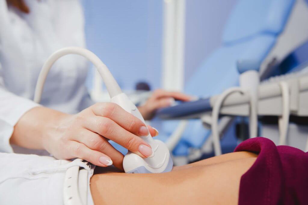
Ultrasound diagnostics (ultrasound) is a classic non-invasive examination method that provides the ultrasound doctor with information about the size, shape and density of the organ under examination, the presence of neoplasms or foreign bodies, as well as focal changes.
MAIN DIRECTIONS OF ULTRASOUND
- Ultrasonography of blood vessels and heart (including head and neck vessels, vessels of extremities) with Doppler effect
- Ultrasonography of abdominal and retroperitoneal organs (liver, gallbladder, pancreas, kidneys, adrenal glands)
- Ultrasonography of the bladder, prostate gland (including through the rectum)
- Gynaecological ultrasound (transabdominal and transvaginal)
- Ultrasonography of mammary glands, thyroid gland, lymph nodes and other superficial structures
- Ultrasonography of joints, soft tissues
Magnetic resonance imaging (MRI)
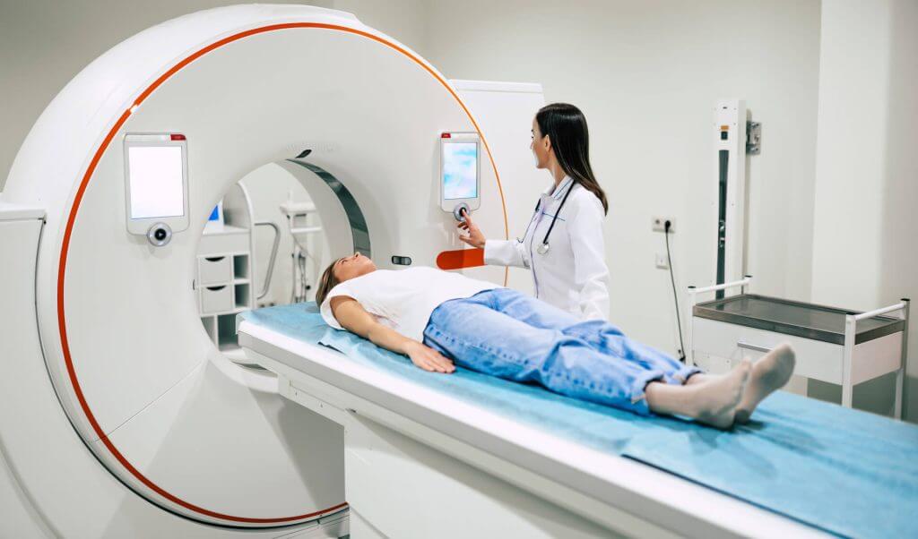
Magnetic resonance imaging (MRI) is a tomographic method of medical imaging and examination of internal organs and tissues using the physical phenomenon of nuclear magnetic resonance (NMR). The method is based on the measurement of the electromagnetic response of atomic nuclei, most often hydrogen nuclei, namely their excitation by a certain combination of electromagnetic waves in a constant magnetic field of high intensity.
This method produces high-contrast images of body tissues and is therefore widely used in medicine, in imaging brain, heart, muscle, and tumour tissue, as compared to other medical imaging methods (such as computed tomography or radiography).
MRI is a non-invasive medical examination method that is widely used in medical diagnostics and monitoring the adequacy of a patient’s treatment. Unlike computed tomography and X-rays, this method does not expose the body to ionising radiation. Instead, the image is formed under the influence of a powerful magnetic field and electromagnetic waves with the use of computer processing to obtain clear detail of soft tissues, bones and other internal structures of the body. Contrast agents are often used to enhance image clarity. MRI can diagnose pathological changes that cannot be seen with other medical imaging methods.
Side effects of MRI are unknown, but there are a number of contraindications.
MRI techniques are constantly being improved. New sequences of electromagnetic pulses are being tested to visualise various body structures and new data processing methods are being applied, which allows for a high level of detail, for example, to image brain regions less than 1 mm thick.
Scientists and companies from various industries are involved in improving the MRI procedure. In particular, Facebook has announced that it is working to teach artificial intelligence to read magnetic resonance imaging results faster. It is planned that the procedure time will be reduced from 30 minutes to 3 minutes, which, in turn, will reduce the price of the procedure. To implement the project, the corporation’s research centre received 3 million MRI scans, 10,000 of which were clinical. The images provided are confidential and do not contain any information protected by law. Together with Facebook, the FAIR AI laboratory and the Department of Radiology at NYU School of Medicine are working on the development.
MRI of the brain
MRI of the brain is used in neurosurgery and neurology, which allows for high accuracy in detecting pathology of brain tissue. Compared to CT scan, this method provides higher sensitivity in diagnosing small tumours and better visualisation of the posterior cranial fossa. The resulting image allows for a clear distinction between grey and white matter, which allows for the diagnosis of a number of pathological processes in the central nervous system, including demyelinating diseases, dementia, cerebrovascular diseases, neuroinfections and epilepsy. Since the examination produces multiple images with a few milliseconds interval, it allows to see the brain’s response to various stimuli, to investigate both functional and structural brain abnormalities, and to diagnose most mental disorders. The method is also used in MRI-guided stereotactic surgery – radiosurgery for the treatment of brain tumours, arteriovenous malformations and other diseases requiring surgical treatment.
MRI of the cardiovascular system
MRI of the cardiovascular system complements other imaging methods, such as echocardiography, cardiac CT and nuclear medicine. Its applications include examinations for coronary heart disease, cardiomyopathy, myocarditis, haemochromatosis, congenital heart disease, etc.
MRI of the musculoskeletal system
Diagnostics of diseases of the musculoskeletal system includes MRI of the spine, diagnostics of joint diseases and soft tissue tumours.
Electroencephalography (EEG)
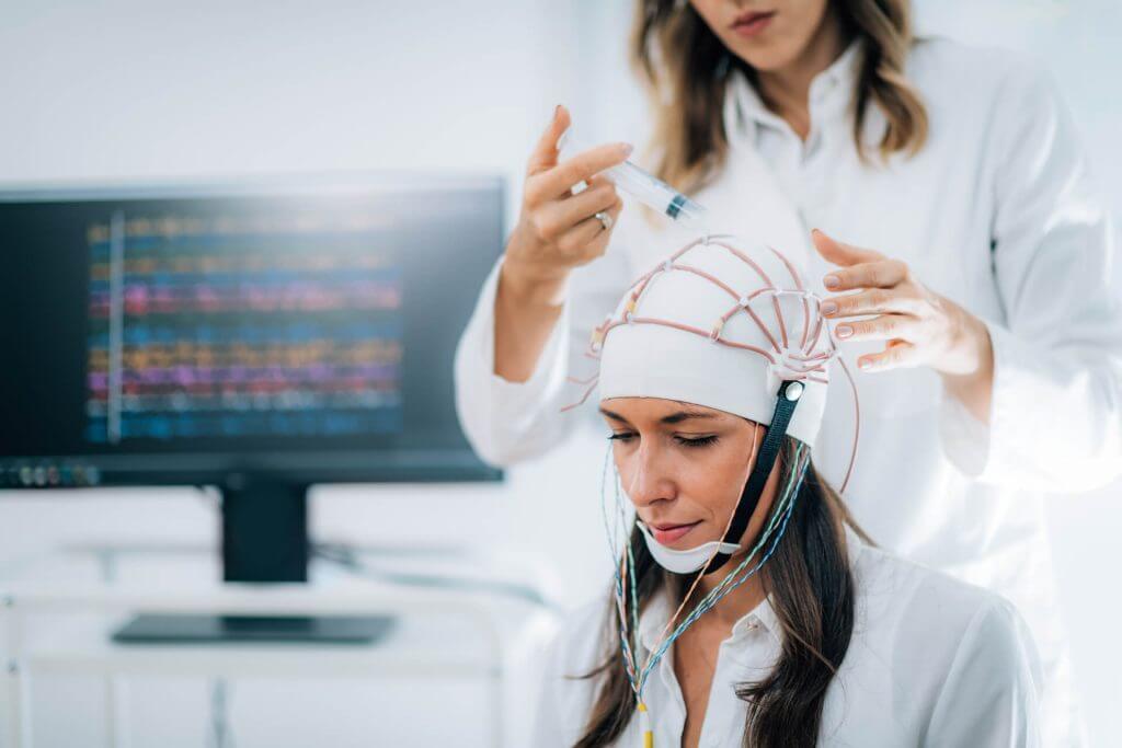
Electroencephalography (EEG) is a method of graphical recording of brain biopotentials, which allows analysing its physiological maturity and condition, the presence of focal lesions, general brain disorders and their nature. It consists in recording and analysing the total bioelectrical activity of the brain – electroencephalogram (EEG). EEG can be taken from a scalp, from the surface of the brain, as well as from deep brain structures. Typically, an electroencephalogram is understood to be a surface recording, i.e., made from the skin. A recording made with electrodes from the surface of the brain is called an electrocorticogram.
The EEG is most commonly used to diagnose epilepsy, which causes abnormal EEG patterns. It is also used to diagnose sleep disorders, depth of anaesthesia, coma, encephalopathy and brain death. The EEG was used as the primary method for diagnosing tumours, stroke and other focal brain disorders, but as high-resolution anatomical images became available through magnetic resonance imaging (MRI) and computed tomography (CT), the use of the EEG declined. Despite its limited resolution, EEG continues to be a valuable tool for research and diagnosis. It is one of the few mobile methods available and has a temporal resolution in the millisecond range, which is not possible with CT, PET or MRI.
Examination Cost (Price)
| Cost of examination according to the clinic’s price list |